Best of the
Best
Editors' picks and our top buying guides


Best of the
Best
Editors' picks and our top buying guides
Latest

Yes, There's a Right Way to Clean and Maintain Your Enameled Cookware. Here's How
27 minutes ago
iOS 17.5 Is Almost Here, but Don't Miss These iOS 17.4 Features
27 minutes ago
Snag 2 Anker USB-C Fast Chargers and Cables for Only $13 With Amazon Prime
42 minutes ago
Best CD Rates Today, April 26, 2024: High APYs Won't Last Forever
57 minutes ago
Best Savings Rates Today, April 26, 2024: Save More With One of These High-Yield Savings Accounts
1 hour ago
Shop Incredible TV Deals at Best Buy This Weekend Only
1 hour ago
Best Buy Launches Huge 3-Day Sale on Top Tech and Major Appliances
1 hour ago
Best Internet Providers for Gaming in 2024
4 hours ago
Best 55-Inch TVs for 2024: Hisense, Samsung and More
7 hours ago
Tesla Powerwall 3 vs. Tesla Powerwall 2: Does Newer Mean Better?
7 hours ago
Today's Wordle Hints and Answer: Help for April 26, #1042
7 hours ago
Best Firm Mattress of 2024
8 hours agoFCC Approves T-Mobile's Deal to Purchase Mint Mobile
8 hours ago
Xbox Will Start Deleting Old Captures on May 30: Here's How to Save Them
9 hours ago
Best Budget 3D Printer of 2024
9 hours agoMore to Explore
Reviews, advice and more from CNET's experts.


Get the best price on everything CNET Shopping helps you get the best prices on your favorite products. Get promo codes and discounts with a single click.
Add to Chrome - it's free!
Our Expertise
Expertise Lindsey Turrentine is executive vice president for content and audience. She has helped shape digital media since digital media was born.
0357911176
02468104
024681024
Featured in
Tech


Upgrade your inbox
Get CNET Insider
From talking fridges to iPhones, our experts are here to help make the world a little less complicated.
Featured in
Money


Crossing the Broadband Divide
Millions of Americans lack access to high-speed internet. Here's how to fix that.


Featured in
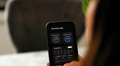
Energy and Utilities


Deep Dives
Immerse yourself in our in-depth stories.


Get the best price on everything CNET Shopping helps you get the best prices on your favorite products. Get promo codes and discounts with a single click.
Add to Chrome - it's free!
Featured in
Internet


Sleep Through the Night
Get the best sleep of your life with our expert tips.


Get the best price on everything CNET Shopping helps you get the best prices on your favorite products. Get promo codes and discounts with a single click.
Add to Chrome - it's free!
Tech Tips
Get the most out of your phone with this expert advice.


Get the best price on everything CNET Shopping helps you get the best prices on your favorite products. Get promo codes and discounts with a single click.
Add to Chrome - it's free!

Featured in
Home


Living Off Grid
CNET's Eric Mack has lived off the grid for over three years. Here's what he learned.